We combine a growing portfolio of ocular disease models with specialized ocular dosing techniques and imaging methods to comprehensively evaluate PK/PD, distribution and adverse effects of novel ocular therapies and devices.
The Biomere team has validated several ocular disease models using state-of-the-art techniques including in-life imaging and terminal imaging and tissue collections. The team focuses on evaluating PK/PD characteristics and early safety profiles of novel therapies to treat ocular diseases. The Biomere site in the US has a variety of ocular models under development and has interest in collaborative model development on new and unique ocular models. Our combined preclinical and clinical research experience distinctively positions us to support our clients’ preclinical ophthalmic needs.
Dosing Techniques
The Biomere ocular team has deep expertise in a range of dosing techniques in mouse, rat, rabbit and primate models. Most of the dosing methods are applicable to large and small animal species, and include the following local and systemic methods:
Topical eye
Intravitreal injection
Subretinal injection
Subconjunctival injection
Intracameral injection
Systemic delivery including oral, subcutaneous, intraperitoneal, intratracheal, and intravenous
Suprachoroidal dosing is limited to rabbit and primate models.
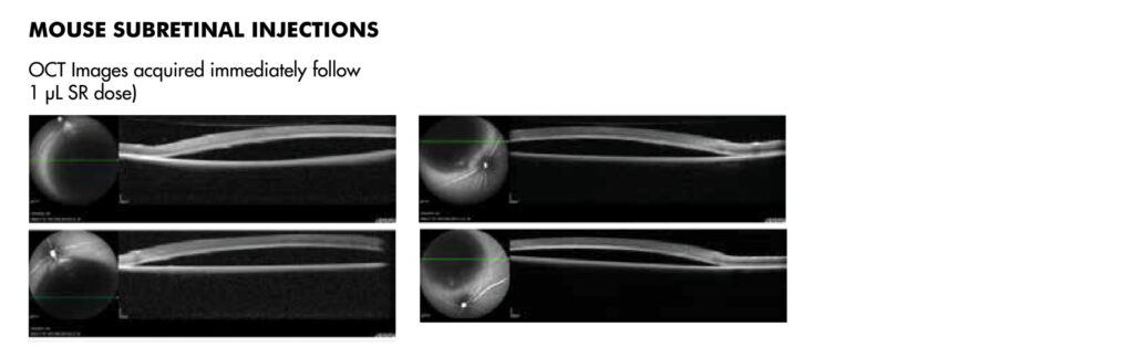
Figure 1: OCT images of mouse eye post sub-retinal injection of 1 microliter SR
On This Page
Ocular Disease Models
The Biomere site in Worcester has validated the following ocular disease models for preclinical studies, and is continuously developing and validating new disease models:
An ocular drug delivery system is a drug formulation that is designed to deliver a therapeutic to a specific part of the eye. A common example of an ocular-specific drug delivery is topically applied eye drops, or an injection into the retina (subretinal) or vitreous humor (intravitreal). Given the complex tissue architecture of the eye, it is very important to perform preclinical testing of the route of administration in relevant animal models, such as rabbits. Ocular experts at Biomere have expertise in multiple routes of ocular drug delivery, including subretinal, intravitreal, subconjunctival and topical to assess ADME characteristics, including distribution and clearance, as well as potential adverse effects, such as acute toxicity, inflammation responses and tissue damage.
What is ocular drug toxicity?
Ocular drug toxicity is an adverse reaction to a therapy that is administered systemically or locally to the eye tissues. Ocular toxicity can also be caused by exposure to organic chemicals, including acids, solvents, etc., which can splash into the eye and damage tissues. The most well-known ocular drug toxicity is damage or inflammation to the conjunctiva, caused by direct exposure. Because the eye has an extensive blood vessel network (vasculature), systemic therapies can cause ocular drug toxicity, especially when the drug can pass through the blood-retina barrier (BRB). Similar to other organ-specific toxicities, ocular drug toxicity can range from mild to severe, and be acute or chronic. Evaluating ocular toxicity in preclinical models is a critical part of early toxicology studies in rodent or large animal models. Once a therapy is administered via a systemic or ocular tissue-specific route, the adverse impact of the therapy can be evaluated using imaging, histo-pathology or changes in gene expression profiles.
What types of drugs are administered to eye tissues?
Many different drug types are administered directly into the eye. The easiest route of administration is topical, where therapies formulated as eye drops are applied directly to the eye surface. This is the most common route for anti-allergy eye drops; glaucoma medications to reduce intraocular pressure; antibiotics to combat infections; and steroids to control inflammation. Topical application is an easy process that does not require medical intervention. However, other ocular routes of administration, such as intravitreal or subconjunctival require a medical professional to administer the drug. Intravitreal injections deliver the therapy directly into the vitreous humor (eyeball), and is one way to deliver therapies for wet AMD or acute macular degeneration caused by the development of abnormal vasculature that damages the macula or central retina. Sub-conjuctival injections are a preferred route to delivering antibiotics that target corneal infections.